Alternatives to Animal Testing, Experimentation and Dissection Articles from All-Creatures.org
Human Lung Chip models radiation-induced lung injury
From CAARE Citizens for Alternatives to Animal Research
December 2023
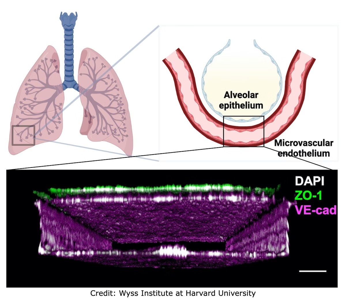
This new human-relevant model combines a Lung Alveolus chip previously developed by Wyss with lung capillary cells, thereby creating an alveolar-capillary interface. Scientists induced radiation damage in the model and found that the resulting changes mimicked actual human lungs, of course, no ANIMALS are used in the process.
The Wyss Institute for Biologically Inspired Engineering at Harvard
University and Boston Childrens Hospital have developed an in-vitro
model of the human lung that can accurately model radiation-induced
lung injury (RILI). RILI can occur following excessive radiation
exposure from nuclear accidents or in some patients receiving
radiation therapy.
Animal experiments do not accurately recapitulate key details of
human lungs, are costly, and pose ethical concerns. Radiation
exposure in animals leads to lung injury, including pulmonary edema,
a painful condition that causes fluid to build up in the lungs
making breathing painful and difficult.
This new human-relevant model combines a Lung Alveolus chip
previously developed by Wyss with lung capillary cells, thereby
creating an alveolar-capillary interface. Scientists induced
radiation damage in the model and found that the resulting changes
mimicked actual human lungs.
They then used the model to test two
drugs to demonstrate that it can be effectively used to test
potential radiation treatments.
This model is just one of many that the Wyss Institute has utilized
to study RILI, including bone marrow and intestine organ chips.
Because radiation damage in one organ could impact the entire body,
researchers hope to eventually link multiple organs together through
a microfluidic device, ultimately testing the full impact of
radiation on the human body.
![]()
Return to Alternatives to Animal Testing, Experimentation and Dissection Articles
Follow us on:
- Home Page
- What's New
- Contact Us
- Action Alerts
- Animal Exploitation
- Animal Issues
- Animal Rights Activism
- Archives
- Art & Photography
- Articles Directory
- Bible
- Bible Commentary Blog
- Books
- Campaigns
- Church & Religion
- Discussions
- Events / Podcasts / Media
- Health
- Hosted Web Sites
- Humor
- Kids' Korner
- Letters
- Links
- Nature Studies
- Newsletter
- Poetry & Stories
- Prayers
- Quotations
- Recipes with Photos
- Recipes Without Photos
- Sermons
- Stop Cruelty in Churches
- Video Library
- The Mary T. and Frank L. Hoffman Family Foundation
- Email: [email protected]
A web site sponsored by The Mary T. and Frank L. Hoffman Family Foundation and all-creatures.org
Copyright © 1998-2026 The Mary T. and Frank L. Hoffman Family Foundation. All rights reserved. May be copied only for personal use or by not-for-profit organizations to promote compassionate and responsible living. All copied and reprinted material must contain proper credits and web site link www.all-creatures.org.
Fair Use Notice: This document, and others on our web site, may contain copyrighted material whose use has not been specifically authorized by the copyright owners. We believe that this not-for-profit, educational use on the Web constitutes a fair use of the copyrighted material (as provided for in section 107 of the US Copyright Law). If you wish to use this copyrighted material for purposes of your own that go beyond fair use, you must obtain permission from the copyright owner.
Thank you for visiting all-creatures.org.
